A Virtual World of Palaeontology
Dr Imran Rahman, Natural History Museum, London
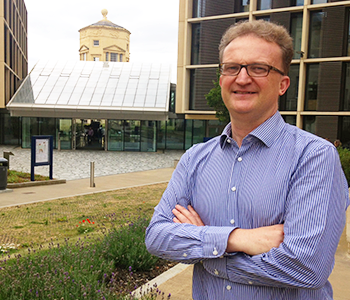
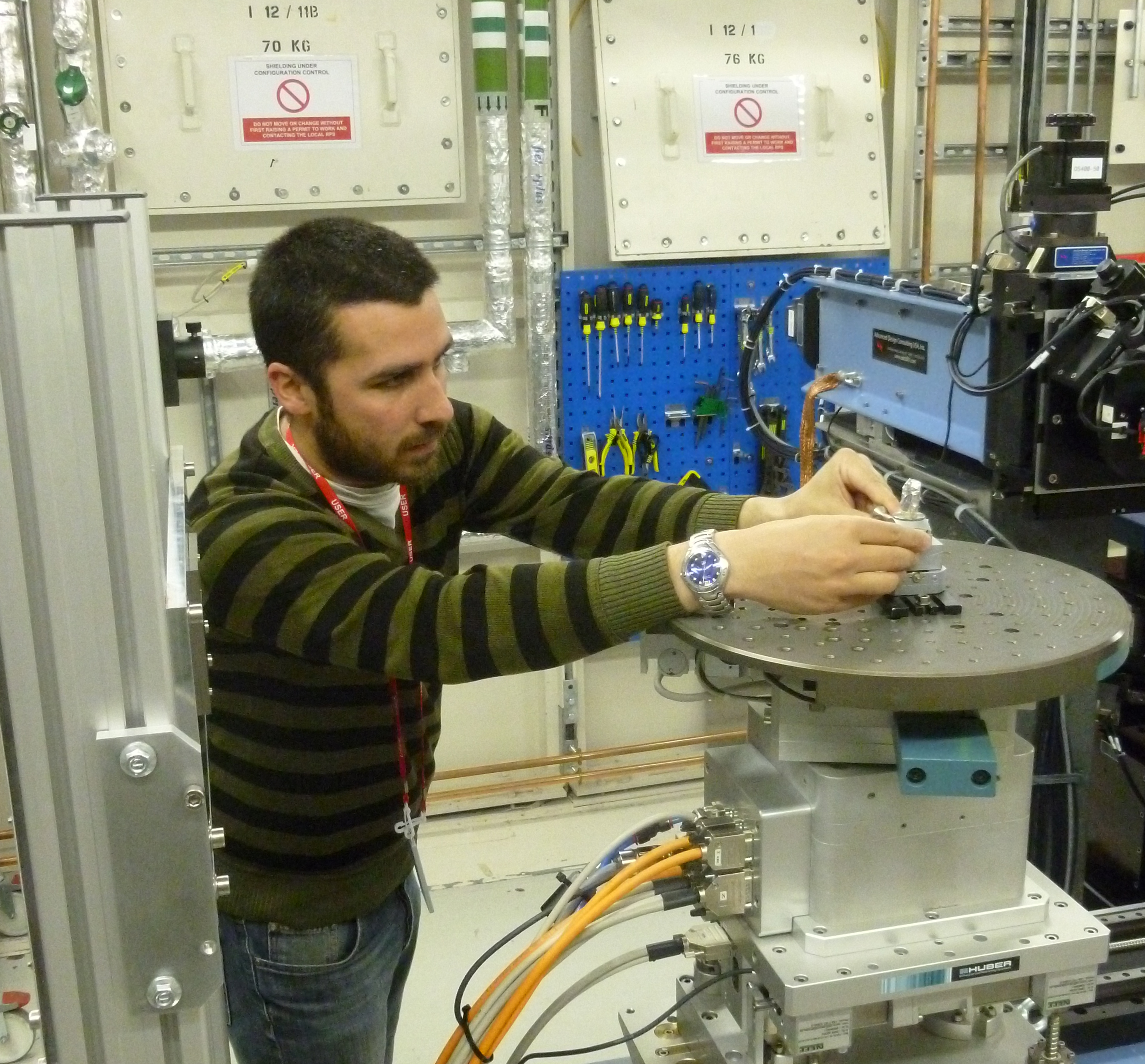
Dr Imran Rahman is a Principal Researcher at the Natural History Museum, London. He is a palaeontologist interested in the origin and early evolution of animals, which dates back over half a billion years to the so-called Cambrian ‘explosion’. His research involves studying the three-dimensional...
Dr Imran Rahman is a Principal Researcher at the Natural History Museum, London. He is a palaeontologist interested in the origin and early evolution of animals, which dates back over half a billion years to the so-called Cambrian ‘explosion’. His research involves studying the three-dimensional and internal structure of exceptionally-preserved fossils using a range of tomographic techniques, including lab-based X-ray micro-tomography, synchrotron tomography and neutron tomography. He is also interested in correlative approaches combining different imaging methods and the application of AI to image segmentation. He currently leads the ‘Phenomics and Advanced Analysis’ research theme at the Natural History Museum, which is focused on the development and application of new tools for imaging and analysis of natural history specimens.